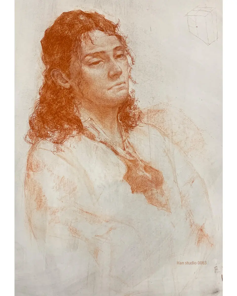
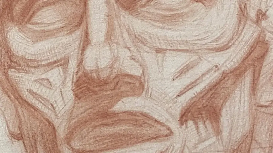
In the process of sketching models, I often accidentally draw too much, like a beard or wrinkles on the face of a teenage girl, and the drawing is too strong to become a big mom. 🙂↕️
Therefore, I am curious about the undulation of the facial muscles, and in the second half of this article, I have organized the characteristics and knowledge of facial muscles, so let's read it together!
After understanding the shape of the face muscles, you can apply it to your portraits to put the light and darkness in the key points.
―――――――――――――――――
Why do I need to learn facial muscles?
The face isThe Main Stage of Emotional PerformanceThe details convey the mood and character of the character.
Painting is not just about depicting surface shapes, suggestsUnderstanding the underlying muscle logic of movement。
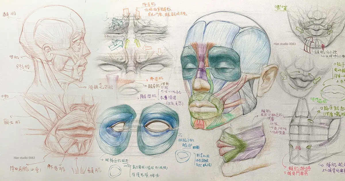
Introduction to the major muscles of the face.
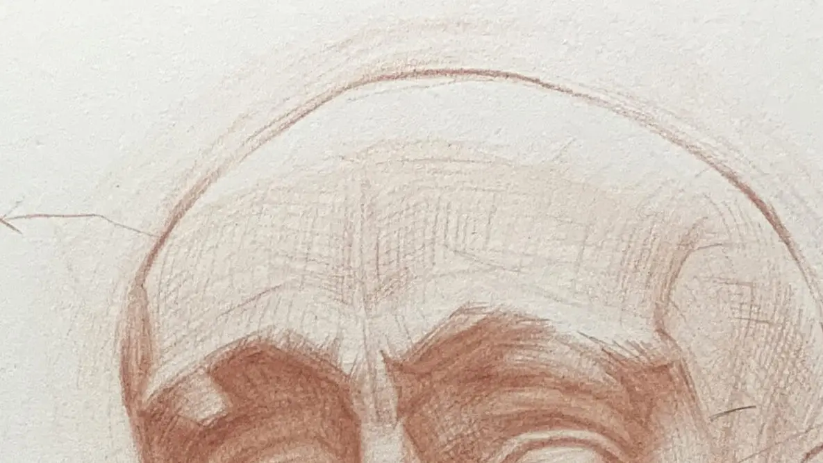
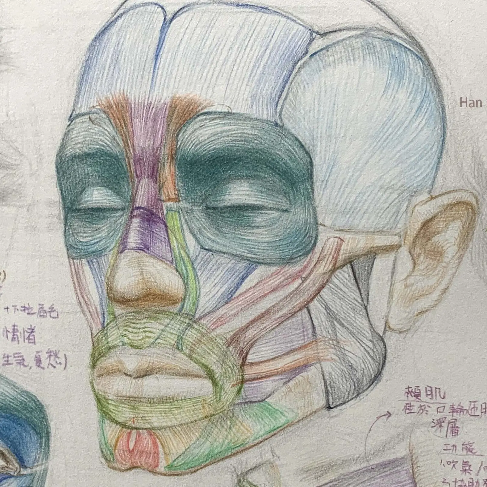
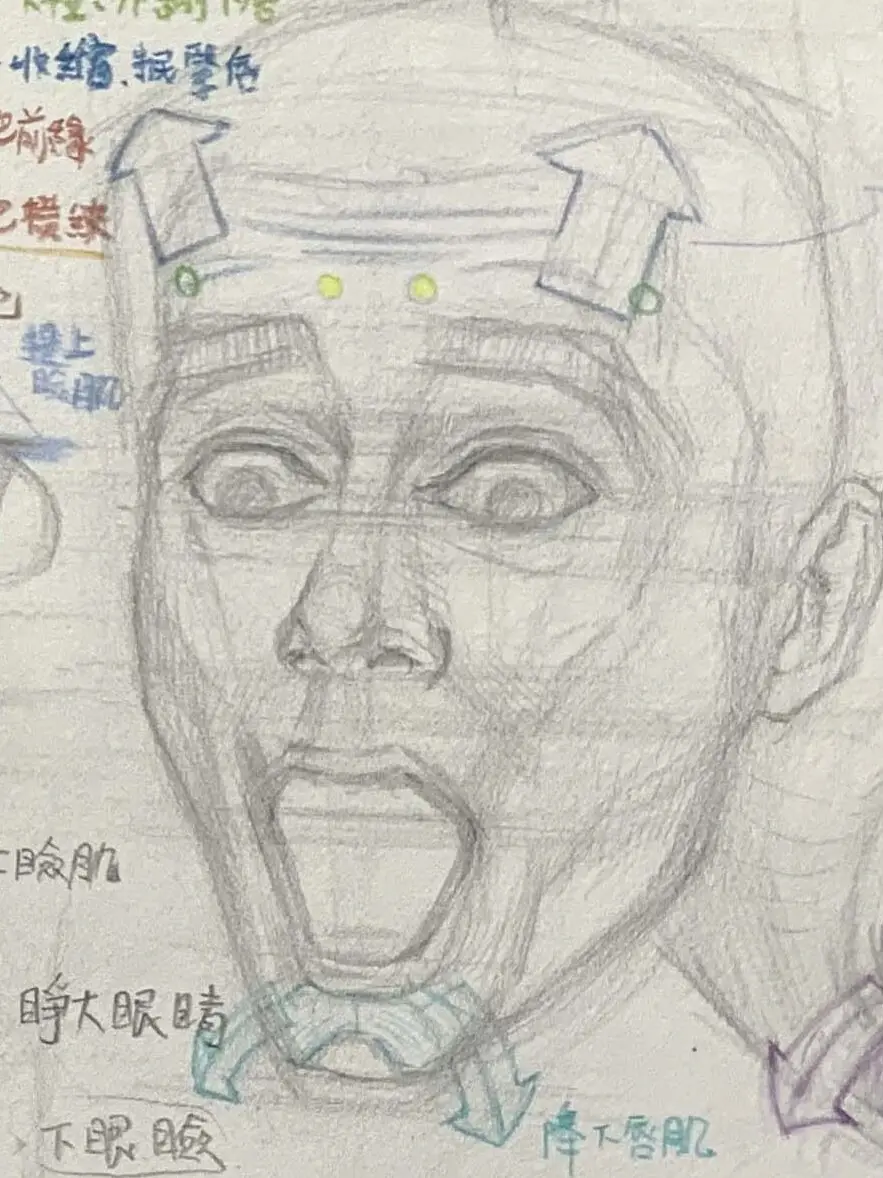
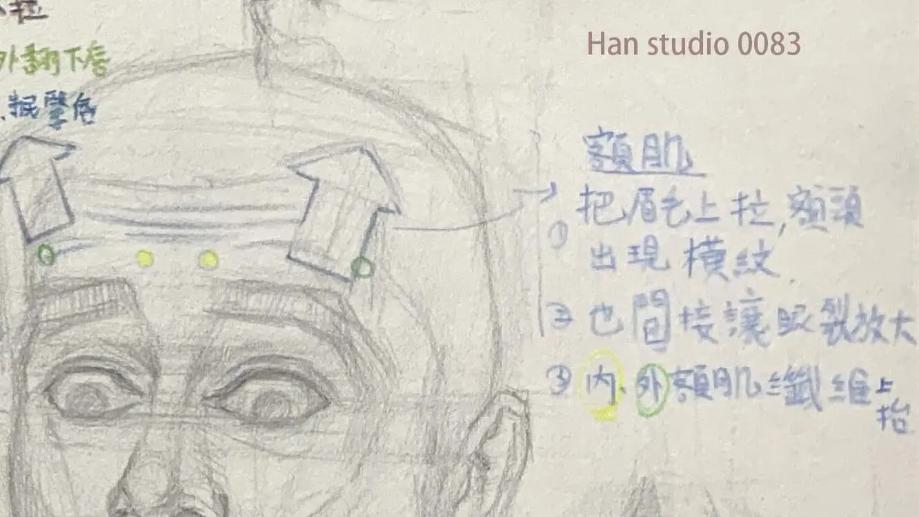
Frontal area
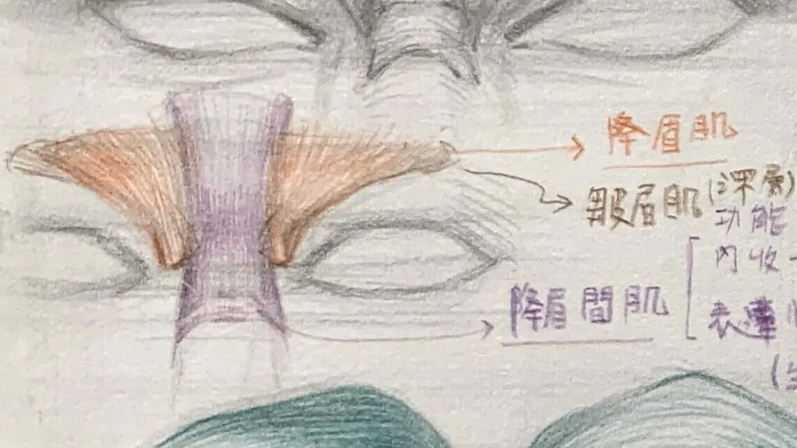
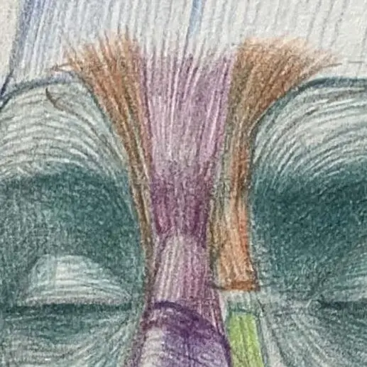
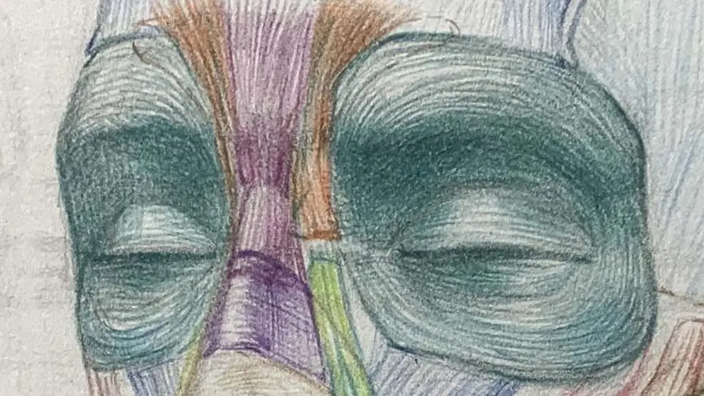
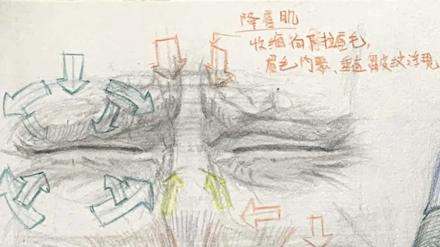
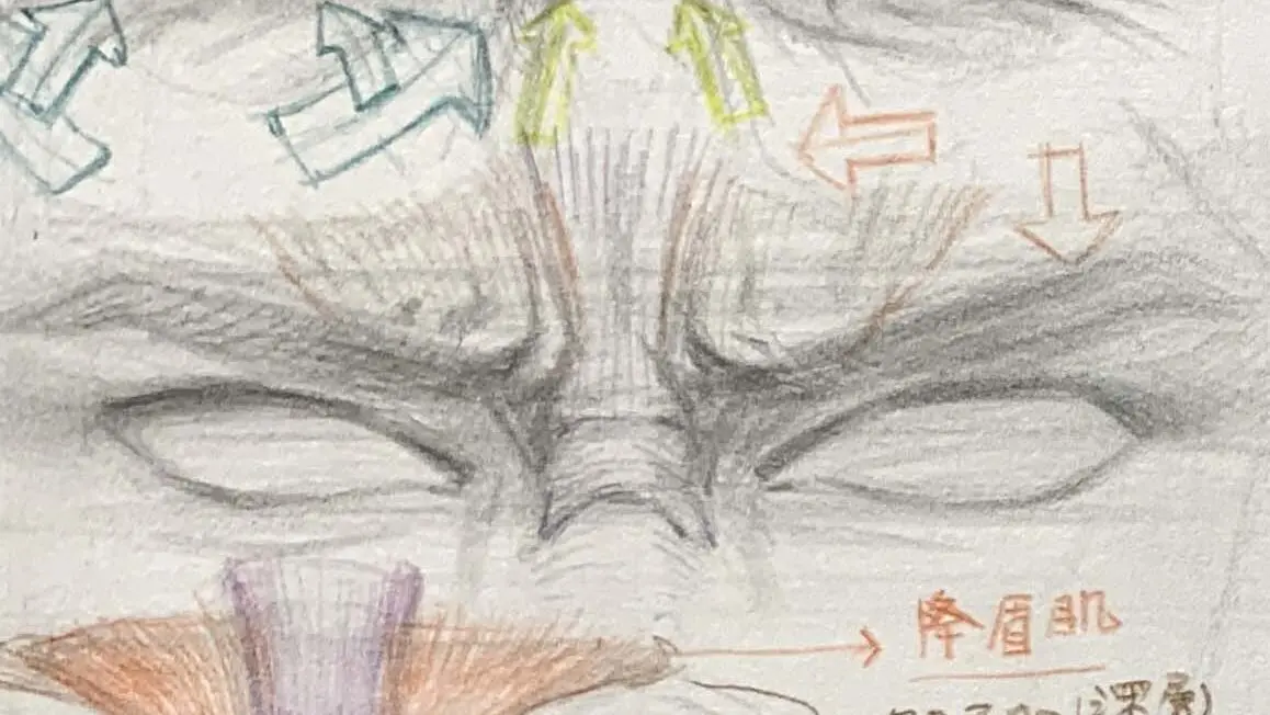
quinine muscle
Shrink to form a raised eyebrow, surprised expression.
Direction of alignment: Vertical alignment is preferred.
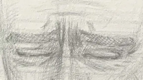
Located on the forehead, it is responsible for raising the eyebrows and frowning. Often used to express surprise (eyes wide open).
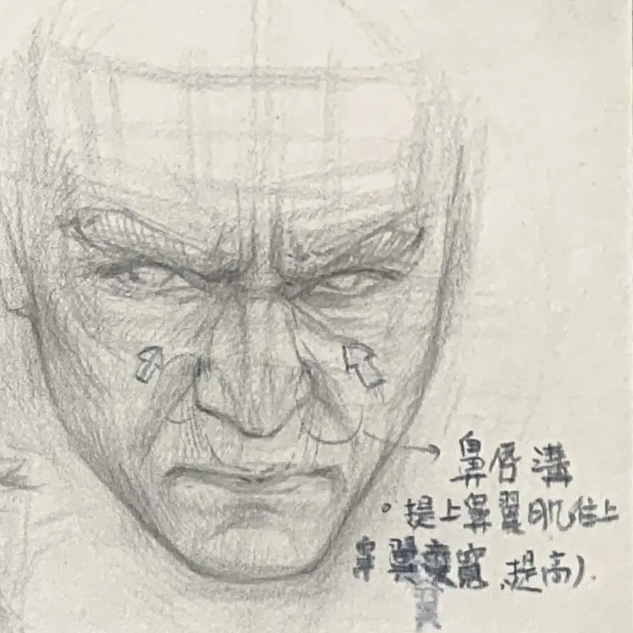
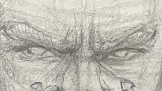
The center of the eyebrows and the root of the nose
Direction of the line: vertical concentration on the center of the eyebrow, radial diffusion.
Around the eyes
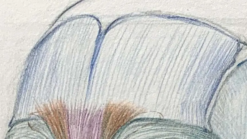
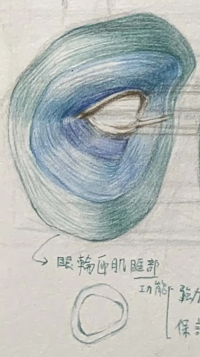
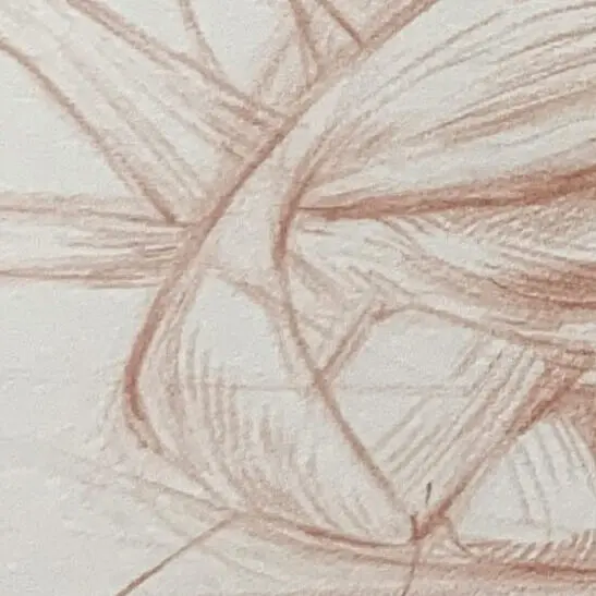
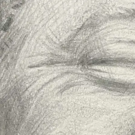
●Orbicularis oculi muscle (sphincter)
Control blinking, closing and squinting.
Direction of alignment: Circular and elliptical around the eyes.
The ring of muscles that surrounds the eyes and is used to close or blink the eyes. When you smile, this area will have natural wrinkles.
The orbicularis oculi muscle can be divided into two main parts:
orbital
Project
Content
Starting point
1. medial border of orbital bone 2. lacrimal sac ridge 3. Periorbital bony rim of the orbit
stop
1. Lateral orbital ligament 2. Intertwine with the eyelid fibers to form a complete ring.
functionality
1. Close your eyes forcibly.(e.g., "squeezing" the eyes, blinking hard) 2. protecting the eyeballs, draining tears
Supporting Graphics
Muscle fibers radiate from the bone edge to the lateral orbital ligament - deep radial wrinkles appear under the brow and on the cheeks when the eyes are drawn in a crowded pattern
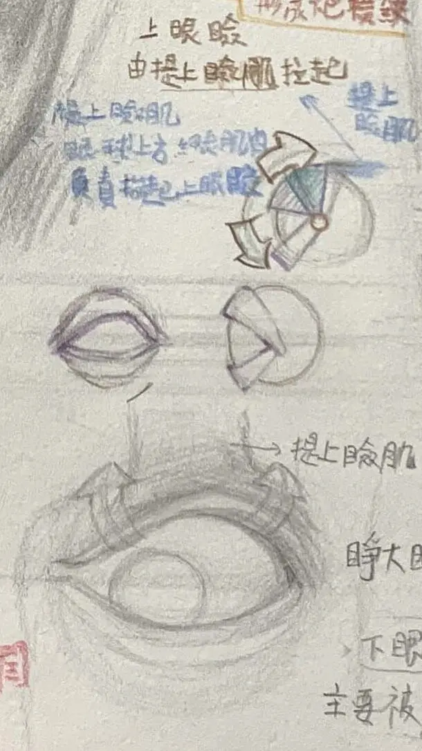
eyelid
Project
Content
Starting point
Near the medial ligament
stop
Lateral Eyelid Ligament
functionality
Soft wink.(natural blinking without wrinkles, maintaining tear film distribution and eyelash cleanliness)
Supporting Graphics
- Located above the upper and lower lids, the muscle fibers run nearly horizontally - when blinking naturally, only this part shrinks without major wrinkles around the eyes.
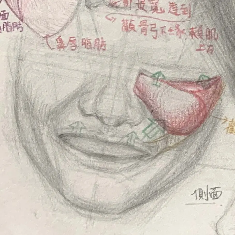
Cheeks and cheekbones
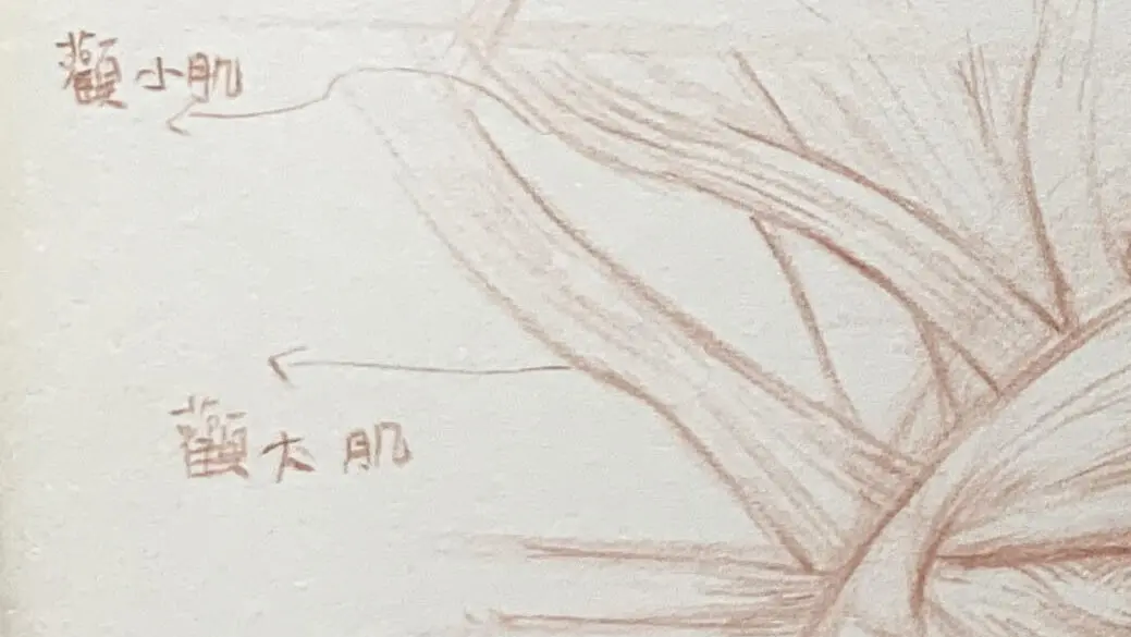
zygomaticus zygomaticus, zygomaticus minor
Produces a smile when the corners of the mouth are raised.
Direction of Line: From the cheekbone to the corner of the mouth diagonally downward.
It extends from the cheekbone to the corners of the mouth and is responsible for pulling up the corners of the mouth to express a smile. This is the key muscle when drawing smile expressions.
Around the mouth
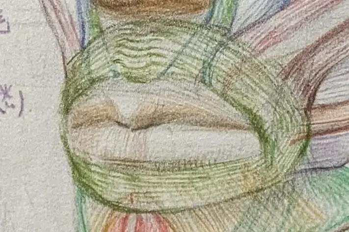
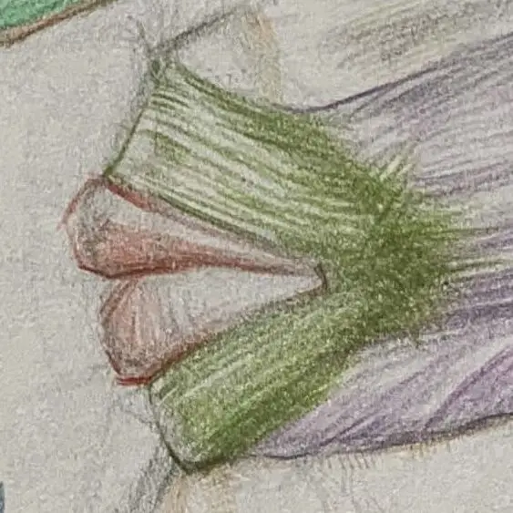
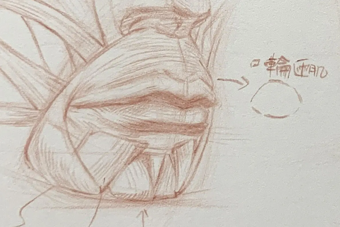
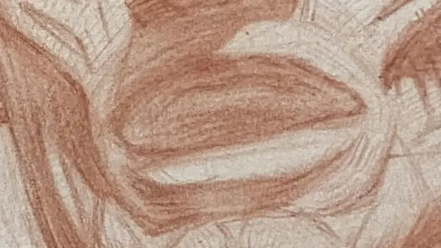
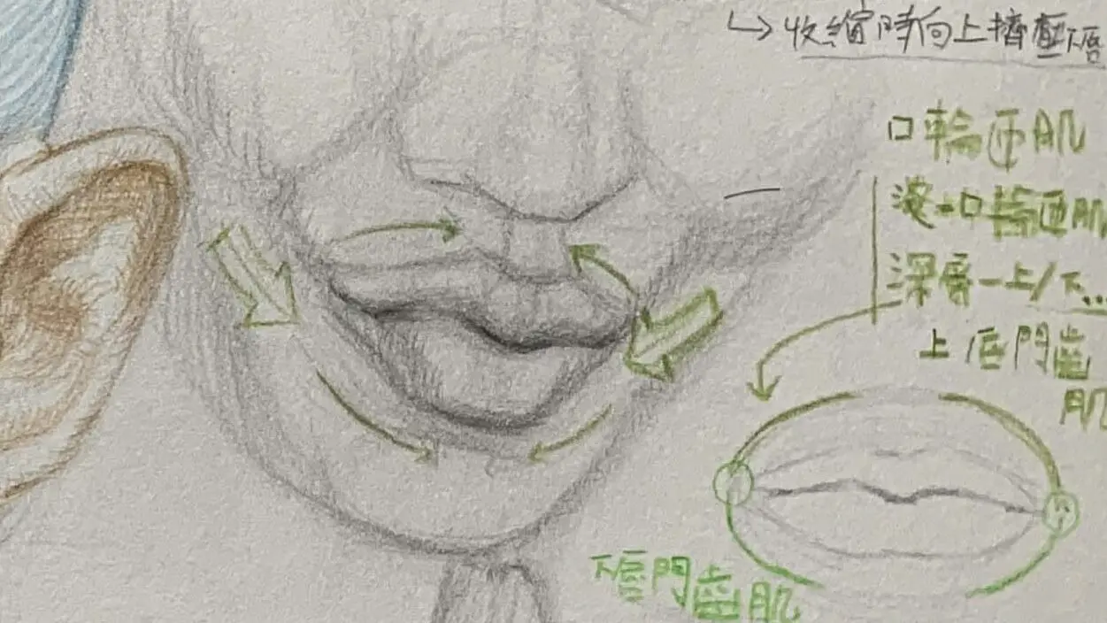
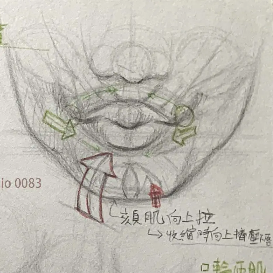
●Oral orbicularis oculi muscle (sphincter)
Pouting and contracted lip expressions.
Direction of rows: Oval shape around the mouth.
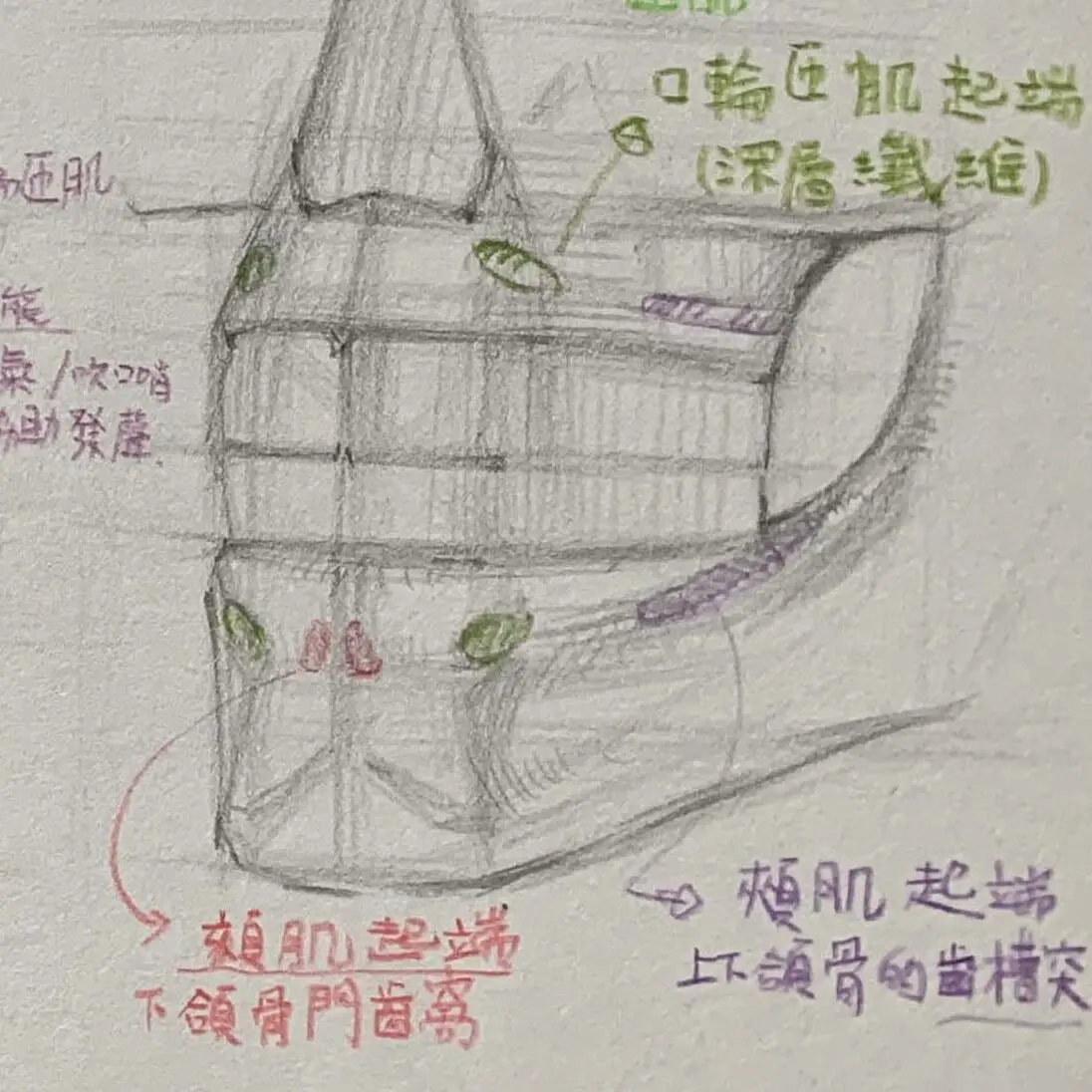
The orbicularis oris muscle is not a single "start → stop" band muscle (usually referred to as "skeletal muscle", i.e., the muscle connected to the skeleton and controlled by consciousness, responsible for body movement), but rather, the orbicularis oculi is a muscle that is used for body movement through theShallow and deep fibers:
DeepThe fibers originate from the grooves of the upper and lower jaw teeth, wrap around the lips of the mouth, and then go to the opposite side;
ShallowThe fibers are formed by the convergence of several peripheral expression muscle bundles in theCorner of the mouth nodule (modiolus)Then wrap it around your lips.
A ring of muscles that surrounds the lips and controls their opening and closing. Used to depict movements such as talking, blowing or pouting.
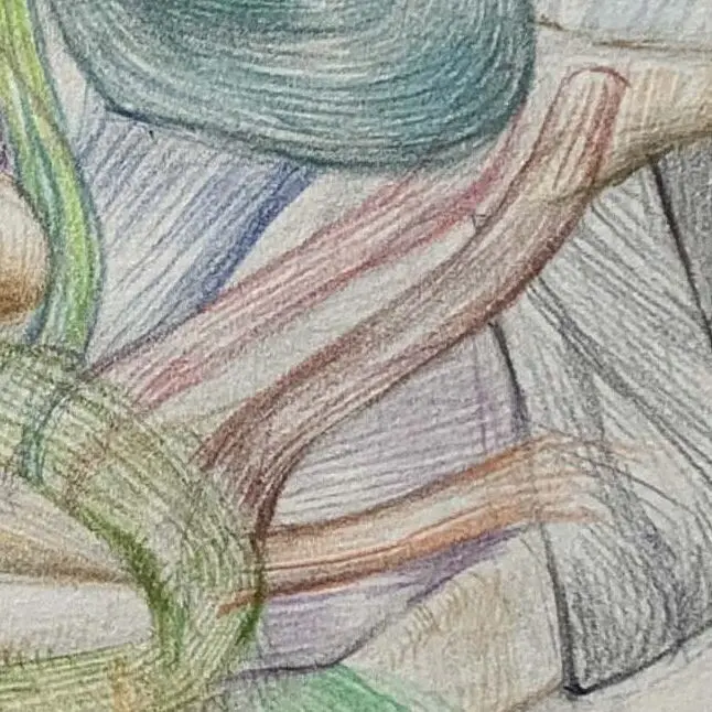
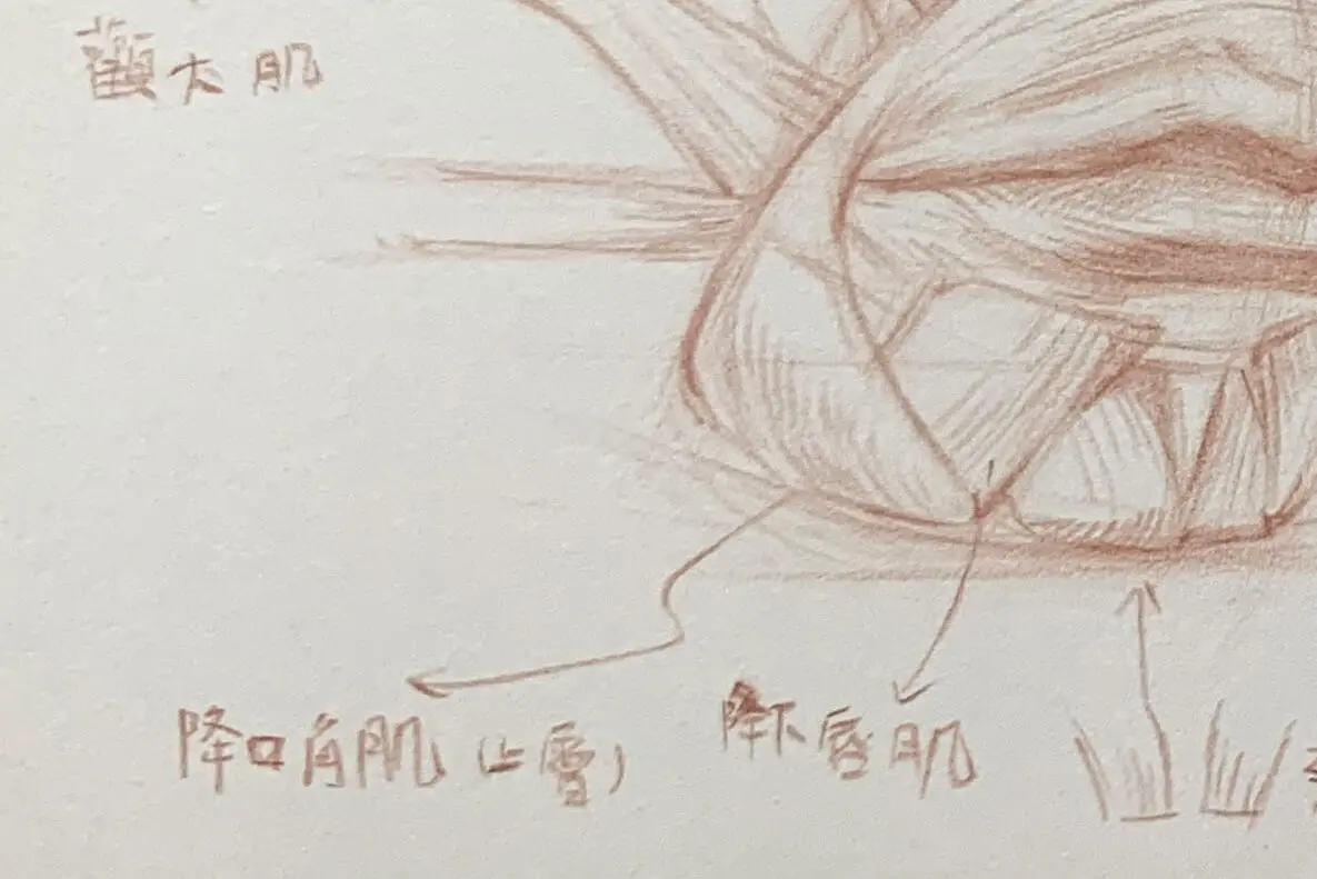
:: Descending corrugator muscle
It is located at the corners of the mouth and pulls down the corners of the mouth to express sadness or dissatisfaction.
The corners of the mouth pull down, creating an expression of sadness or dissatisfaction.
Direction of rows: vertically from the corners of the mouth to the bottom.
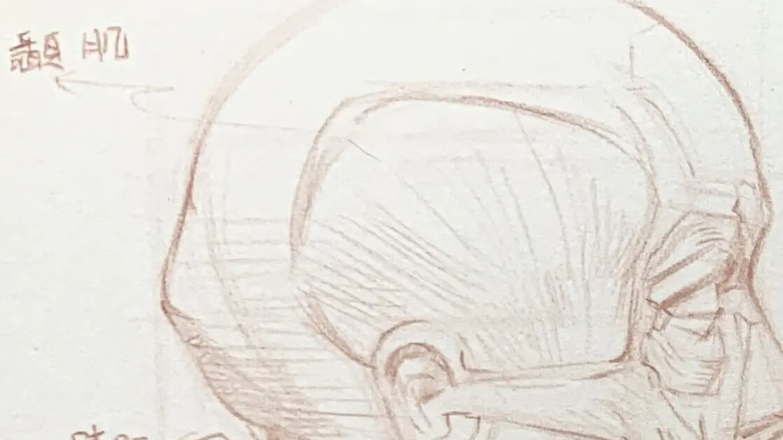
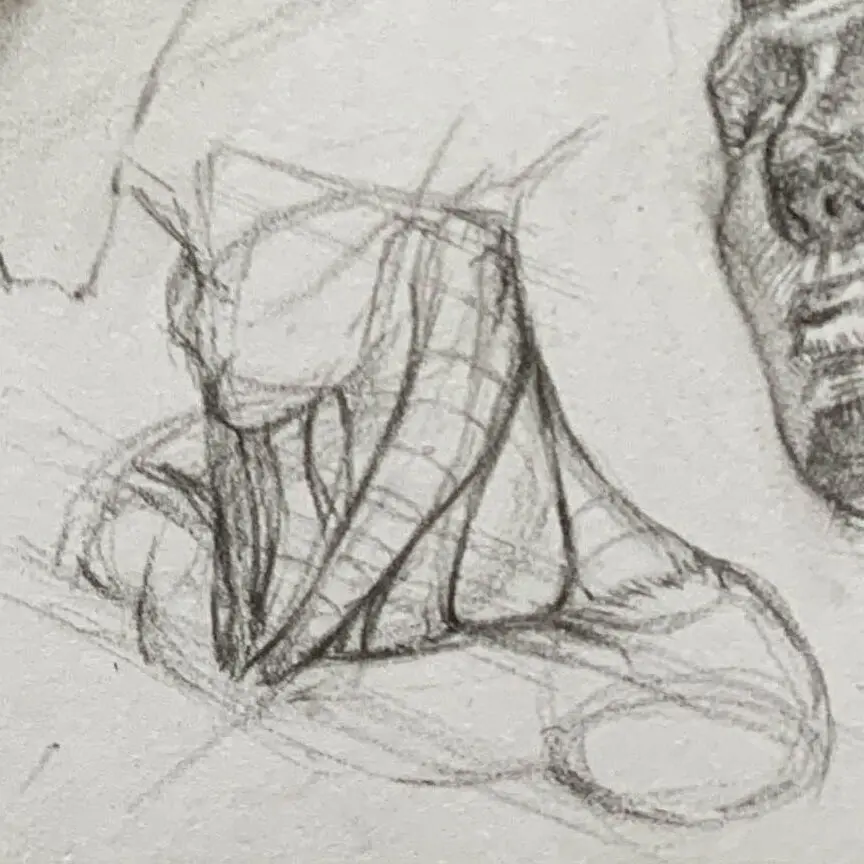
Jaw and side of the face
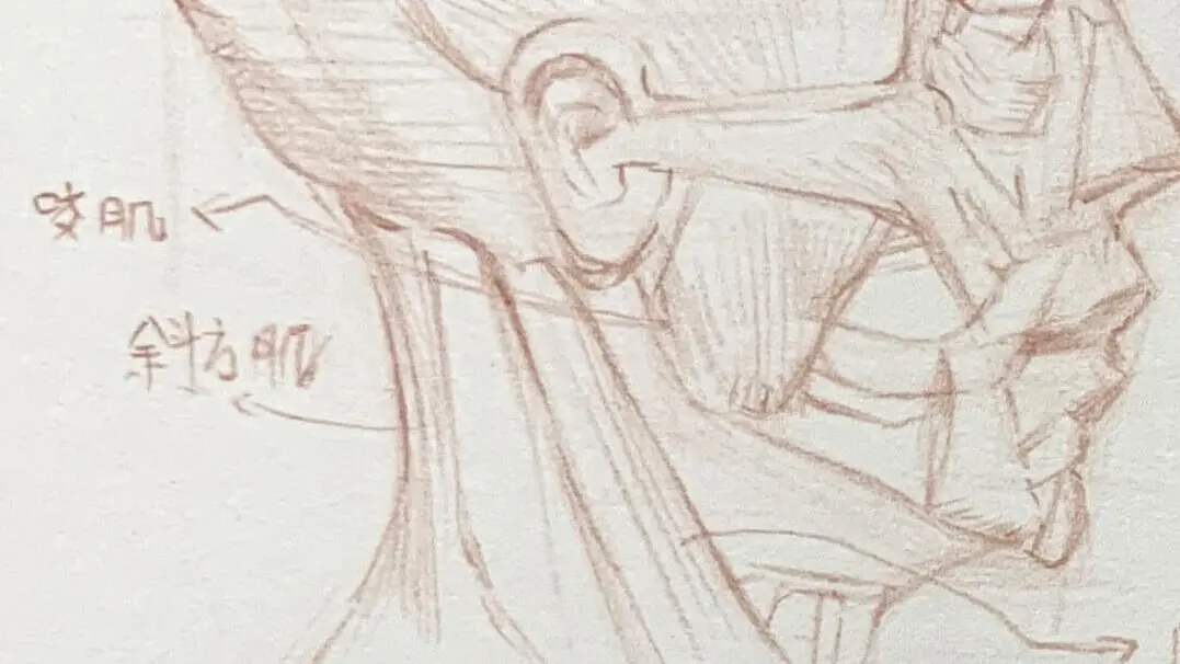
●Bite
A distinctive undulating sensation when chewing, with the side of the cheeks standing out clearly.
Direction of rows: Vertical rows are mostly used to emphasize the three-dimensionality of the muscle bulge.
It is located on the side of the cheek and is responsible for closing the jaw. This is the muscle that expresses chewing or clenching.
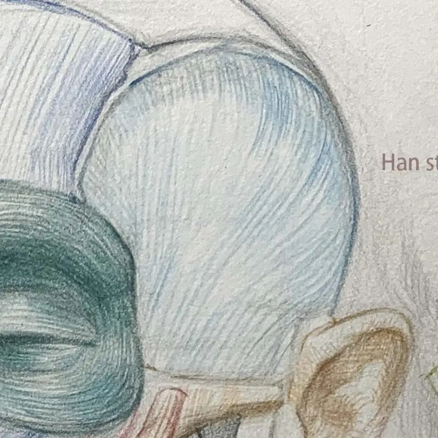
temporal muscle
Position: Located on the side of the skull, it is responsible for controlling the opening and closing of the lower jaw. Characteristics: The temporal muscles are attached to the sides of the skull in an approximate fan shape.
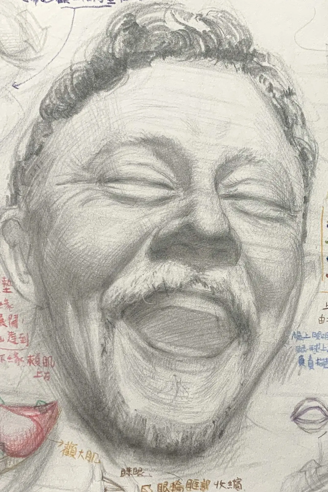
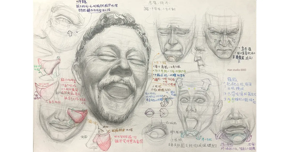
Muscle group coordination and complex expression analysis
Expressions are usually formed by several groups of muscles working together.
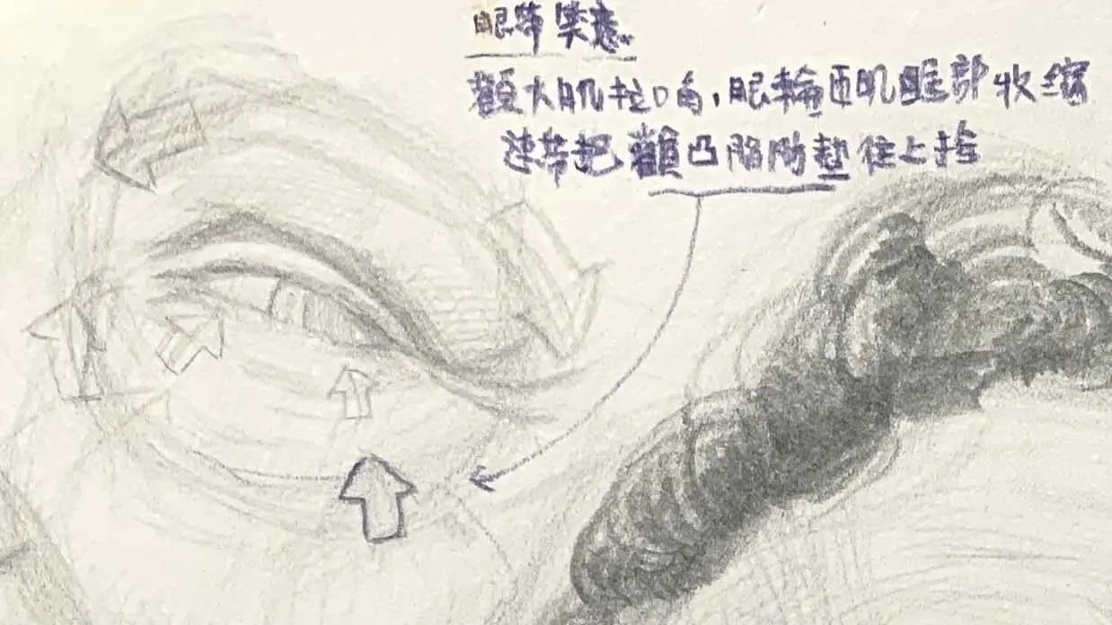
Action: Light, natural blinking/half-closing of the eyes to keep the eyelids against the eyeballs.
In this action: In order to tighten the eye area, the eyelids are first contracted, making the crease smaller.
Orbicularis oculi (orbital)→Crow's feet pull in at the temples and radial creases develop under the eyebrows and on the cheeks.
Action: When the eyes are squeezed forcefully, they are pulled outward from the edge of the orbital bone, resulting in fine lines radiating from the corners of the eyes.
In this action: The large area of the orbital region shrinks, which in turn pulls the skin under the brow, causing wrinkles to appear at the outer corners of the eyes and zygomatic folds.
Expression drawing advice: watch the eyebrowsClip angle change與Direction of forehead crease
periocular muscle group
orbicularis oculi muscle (anatomy): Wraps around the eyes and retracts when eyes are closed or squinting.
eyelid lift: Control of the eyes
Description suggestion: when eyes are closed, the upper eyelid is pressed down and the lower eyelid is bulging out.
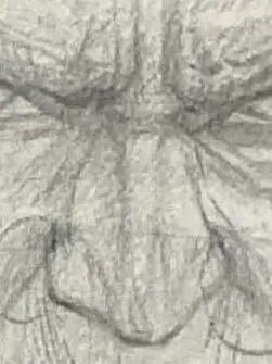
Muscles around the nose
Lowering of the nasal muscles, lifting of the upper lip and nose muscles: Affects nasal tension, wrinkled nose expression
Extendable Supplement:Inhale. Gross. Disgusted.source of expressions
Lips and Cheeks
orbicularis oculi muscle (anatomy): contract the lips to form a rounded shape (e.g., beak, blow)
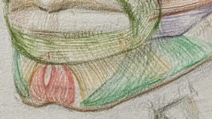
A "pout" is mainly based on the orbicularis oculi muscle.The different layers of fibers are synergistically shrunk, and then combined with theThe chin assisted. The details of the movement are as follows:
Deep orbicularis oris muscle (upper lip incisor muscle)) Fiber Shrinkage
Deep fibers wrap around the inside of the lip, pulling the edge of the lip tighter so that the upper and lower lips come together towards the midline, creating a "slit".
Fiber contraction of the superficial orbicularis oculi muscle (lower lip incisor muscle)
Superficial fibers encircle the outside of the lip, pushing the labrum forward and causing the lip to roll over and arch outward.
Jogging Board
Located in the center of the lower lip, it squeezes the lower lip upward during contraction, enhancing the convexity of the lower lip and the overall protruding effect of the lips.
Slight contraction of the cheek muscles
Helps to stabilize the pressure on the cheeks so that the air pressure in the mouth is more concentrated in front of the lips, resulting in a fuller, more even lip shape.
Lift the corners of the mouth and smile muscles: Lift up the corners of your mouth (happy, polite)
Lowering the corners of the mouth and chin muscles: Sadness, Sagging
Expression Painting Suggestion: Observe MoreCorner of the mouth muscle direction arrows with proportional changes
Jaw and lateral neck muscles
jogging board: Relating to the contraction of the jaw, expressing emotions such as grief and patience.
sternocleidomastoid muscle (anatomy): Although it is a muscle of the neck, it is an important line that expresses tension and vocalization.
Tracing suggestion: exaggerated expression, neck muscles obviously pull out the tension of the line
How to Exercise: From Muscle Figure to Expression Observation
The proposed operation is as follows:
Blank structural drawing of the face layered with muscles (layered like the demo drawing)
Make 3~5 typical emotions: happy/angry/surprised/sad/disgusted.
用The arrows show the direction of contraction of each muscle.The first is to break down the expression into a source of muscle action.
Observe your muscle movements (especially the corners of your mouth and brow) with mirrors/selfies
Advanced Application Suggestions: Linking Dynamics to Character Design
The expression is not static, butSuccessive changes in muscle action。
Extended learning is recommended:
Split "happy" into: smile → laugh → frown and smile
Observe at each stage: which muscles are active and which are passive traction
I hope that this introduction to the structure can help you recognize and understand the knowledge of facial muscles, if there are any subjects you would like to see or drawing methods you would like to know, please feel free to leave a message or private message me, I wish you a happy painting!
―――――――――――――――――
If you want to learn to draw, but don't know how to start, or are interested in understanding what drawing is all about.